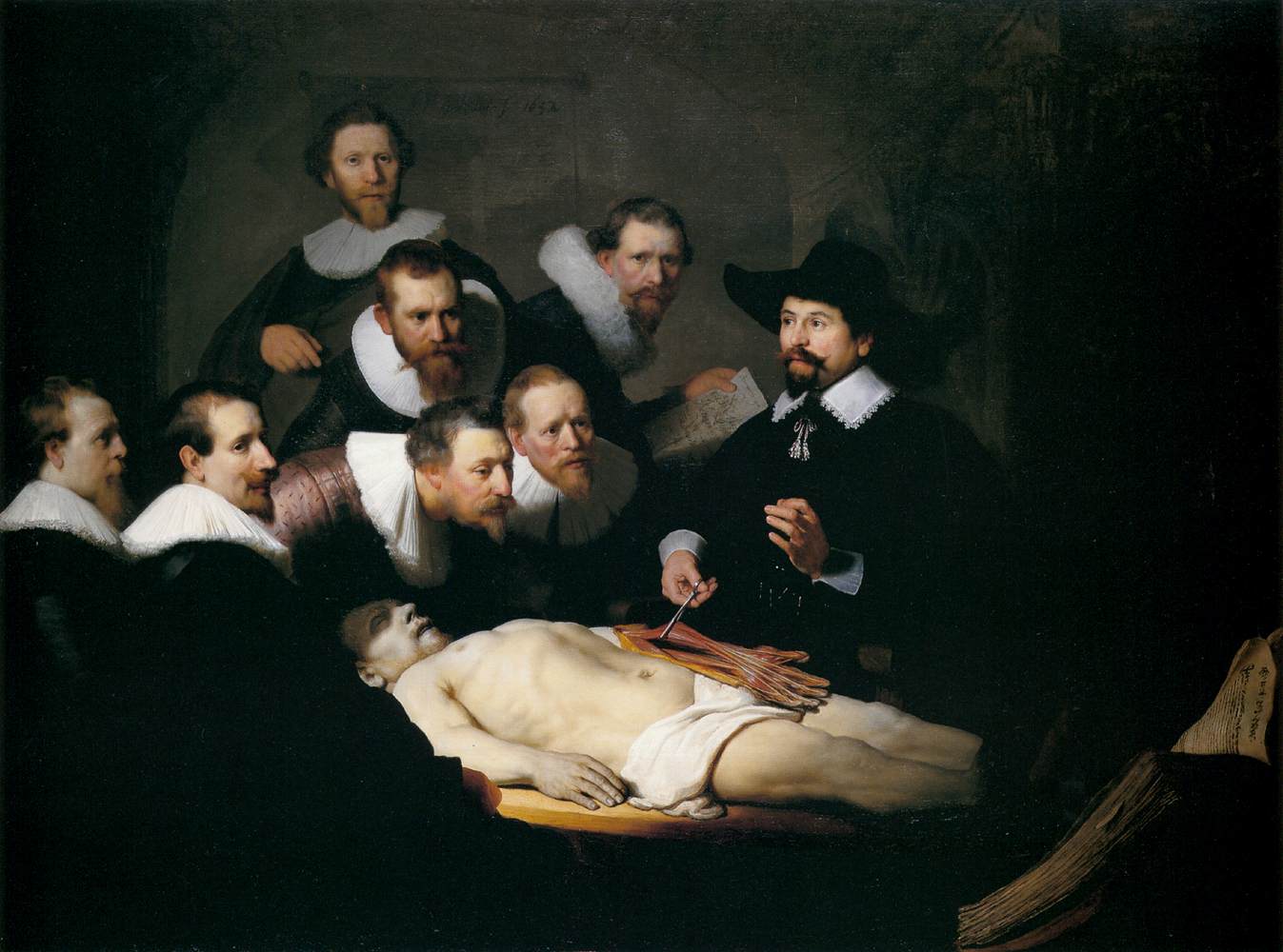
The Anatomy Lesson of Dr. Nicolaes Tulp is a 1632 oil painting by Rembrandt.
Dr. Nicolaes Tulp is pictured explaining the musculature of the arm to medical professionals

Superficial Muscles of the Human Skeleton

The following is a list of muscles you will need to know for class. Included with the name is the origin and insertion and the action the muscle creates. They are organized by location/action.


MUSCLES OF HEAD AND NECK

Facial Muscles - Lateral View       Facial Muscles - Front View       Facial Muscles - Side View

Name Origin and Insertion Action
Epicranius: Frontalis & Occipitalis Frontalis:Forehead to Galea Aponeurotica; Occipitalis:Galea Aponeurotica to Occipital Bone Raises the eyebrows
Orbicularis Oculi Circular muscle around the eyes Blinks and closes eyes
Orbicularis Oris Circular muscle around the mouth Closes and protrudes lips
Buccinator Alveolar processes of mandible and maxilla to skin around mouth Flattens cheek against teeth
Zygomaticus Major and Zygomaticus Minor Zygomatic bone to angle of mouth/orbicularis oris Raises corner of mouth
Platysma From edges of Deltoid and Pectoralis Major muscles upward to lower border of mandible Draws mouth downward
Levator Labii Superioris Maxilla to Orbicularis Oris Raises upper lip
Depressor Labii Inferioris Mandible to Orbicularis Oris Depresses (lowers) lower lip
Risorius Fascia over Parotid (salivary) Gland to skin at angle of mouth Draws angle of mouth laterally, as in grimacing
Mentalis Found in the middle of the body of the mandible between the two mental foramen Pouting
Corrugator Supercilii Above superciliary arch of frontal bone, medial to eyebrows Wrinkles skin of forehead as in frowning

MUSCLES OF MASTICATON

Masseter Maxilla and zygomatic arch to angle and ramus of mandible elevates mandible as in closing mouth
Temporalis Parietal bone to coronoid process of mandible elevates, retracts and laterally moves mandible
Medial Pterygoid Pterygoid process of sphenoid bone to angle and ramus of mandible elevates and protracts (protrudes) mandible and moves mandible from side to side
Lateral Pterygoid Pterygoid process of sphenoid bone to condyle of mandible protracts (protrudes) mandible, depresses mandible, as in opening mouth and moves mandible from side to side

MUSCLES OF THE TONGUE AND NECK

Genioglossus Inside middle of Mandible to tongue depresses tongue
Styloglossus Styloid process of Temporal bone to tongue elevates tongue
Palatoglossus Soft palate to side of tongue elevates posterior tongue/draws soft palate down on tongue
Hyoglossus Hyoid bone to tongue depresses tongue
Digastric Anterior Belly: from Mandible to Hyoid/Posterior Belly: from Hyoid to Mastoid process of Temporal bone elevates Hyoid bone, depresses mandible as in opening the mouth
Stylohyoid Styloid Process of Temporal bone to Hyoid bone elevates Hyoid
Mylohyoid Inside, sides of Mandible to Hyoid bone elevates Hyoid
Geniohyoid Inside, middle of Mandible to Hyoid bone elevates Hyoid Bone
Omohyoid Superior border of Scapula to Hyoid bone depresses Hyoid Bone
Sternohyoid Manubrium of Sternun to Hyoid bone depresses Hyoid Bone
Sternothyroid Manubrium of Sternum to Thyroid Cartilage depresses Thyroid Cartilage
Thyrohyoid Thyroid Cartilage to Hyoid bone elevates Thyroid Cartilage
Sternocleidomastoid Sternum and Clavicle to Mastoid Process of Temporal bone laterally rotate head
Anterior Scalene C-3 thru C-6 to first rib elevates first rib during deep inhalation
Middle Scalene C-2 thru C-7 to first rib elevates first rib during deep inhalation
Posterior Scalene C-4 thru C-6 to second rib elevates second rib during deep inhalation


MUSCLES OF THE THORAX, PECTORAL GIRDLE AND ARMS

TRUNK MUSCLES I       TRUNK MUSCLES II       TRUNK MUSCLES III

TRUNK MUSCLES IV       TRUNK MUSCLES V       TRUNK MUSCLES VI

Subclavius Under 1st rib to clavicle depresses clavicle
Trapezius Occipital bone, C-7, all thoracic vertebrae to spine of scapula, acromion, lateral 1/3 of clavicle upward rotate and adducts the scapula
Latissimus Dorsi All lumbar vert., sacrum, coccyx and posterior iliac crest to intertubercular sulcus of the humerus extends, adducts and medially rotates humerus at the shoulder
Supraspinatus Supraspinous fossa of scapula to greater tubercle of the humerus abducting arm at shoulder
Infraspinatus Infraspinous fossa of scapula to greater tubercle of humerus laterally rotates arm at shoulder
Subscapularis Subscapular fossa of scapula to lesser tubercle of the humerus medially rotates arm at shoulder
Teres Major Inferior angle of the scapula to lesser tubercle of humerus extends arm at shoulder
Teres Minor Axillary border of the scapula to greater tubercle of humerus extends arm at shoulder
Rhomboideus Major T-2 through T-5 to vertebral border of scapula below the spine of the scapula adducts the scapula
Rhomboideus Minor C-7 through T-1 to vertebral border of scapula at the level of the spine of the scapula adducts the scapula
Levator Scapulae C-1 through C-4 to vertebral border of scapula above the spine of the scapula elevates the scapula
Pectoralis Major Medial 1/2 of Clavicle, Sternum and Costal Cartilages to the Greater Tubercle of the Humerus Adducts humerus; pulls arm across chest, medial rotation of arm
Pectoralis Minor Ribs 3-5 to the Coracoid Process of the Scapula Abducts the Scapula and rotates it downward
Rectus Abdominis Pubic crest and pubic symphysis to xiphoid process/costal cartilages. (Note: Tendinous Intersections (Inscriptions)) Flexes vertebral column/compresses abdomen
External Abdominal Oblique Ribs 5-12 to iliac crest and linea alba Flexes vertebral column
Internal Abdominal Oblique Iliac crest to costal cartilages of ribs 7-12 and the linea alba Flexes vertebral column
Transversus Abdominis Iliac crest/lumbar vert./costal cartilages of ribs 5-10 to the xiphoid process, linea alba and pubis compresses abdomen
Diaphragm Circular muscle dividing the thoracic and abdominal cavities/Inserts onto 'Central Tendon' contraction causes it to flatten out, pulling down the lungs for inhalation
External Intercostals Inferior border of rib above to superior border of rib below contraction elevates ribs for inhalation
Internal Intercostals Superior border of rib below to inferior border of rib above contraction draws ribs together for exhalation
Serratus Anterior First 9 ribs to ventral, vertebral border of scapula abducts scapula
Deltoid Spine of scapula, acromion and lateral 1/3 of clavicle to deltoid tuberosity of humerus abduct arm at shoulder
Coracobrachialis Coracoid process of scapula to middle, medial humerus/opposite deltoid tuberosity adducts arm at shoulder

ARM AND SHOULDER:

Biceps Brachii-Long Head Supraglenoid Tubercle of Scapula to radial tuberosity Flexes elbow and supinates hand
Biceps Brachii-Short Head Coracoid Process of Scapula to radial tuberosity Flexes elbow and supinates hand
Triceps Brachii-Long Head Infraglenoid Tubercle of the Scapula to the olecranon process of ulna Extends elbow
Triceps Brachii-Lateral Head Posterior Humerus above the radial groove to the olecranon process of ulna Extends elbow
Triceps Brachii-Medial Head Posterior Humerus below the radial groove to the olecranon process of ulna Extends elbow
Anconeus Lateral epicondyle of the humerus to olecranon process extends forearm at elbow
Pronator Teres Medial epicondyle of humerus to lateral surface of the radius Rotates arm medially; Pronation
Supinator Lateral epicondyle of humerus to lateral surface of the radius Rotates forearm laterally; Supination
Brachialis Anterior, distal 1/2 of the Humerus to the coronoid process of the ulna Flex the Elbow
Brachioradialis Lateral Supracondylar ridge of Humerus to the styloid process of the radius Flexes forearm at elbow
Flexor Carpi Radialis Medial epicondyle of the humerus to second and third metacarpals Flexes wrist
Flexor Carpi Ulnaris Medial epicondyle of the humerus to pisiform, hamate and fifth metacarpals Flexes wrist
Palmaris Longus Medial epicondyle of humerus to flexor retinaculum (palmar aponeurosis) Flexes wrist
Extensor Carpi Ulnaris Lateral epicondyle of the humerus to the fifth metacarpal Extends wrist
Extensor Carpi Radialis Longus Lateral epicondyle of the humerus to second metacarpal Extends hand at the wrist
Extensor Carpi Radialis Brevis Lateral epicondyle of the humerus to third metacarpal Extends the hand at the wrist
Extensor Digitorum Lateral epicondyle of the humerus to second to fifth phalanges Extends fingers/wrist
Extensor Digiti Minimi Lateral epicondyle of the humerus/tendon of extensor digitorum to the fifth phalanx Extends little finger
Intrinsic Muscles of the Hand Thenar lateral aspect of palm (thumb side of palm)
Intrinsic Muscles of the Hand Hypothenar medial aspect of palm (little finger side of palm
Intrinsic Muscles of the Hand Intermediate (Midpalmar) middle of palm
Erector Spinae iliocostalis group lateral aspect of spine
Erector Spinae longissimus group intermediate (in the middle)
Erector Spinae spinalis group medial aspect of spine


MUSCLES OF THE PELVIC GIRDLE AND THE LEGS

(Iliopsoas) Psoas Major lumbar vertebrae to lesser trochanter of the femur flex thigh at hip
(Iliopsoas) Iliacus iliac fossa to lesser trochanter flex thigh at hip
Quadratus Femoris ischial tuberosity to intertrochanteric crest laterally rotates at hip joint
Adductor Longus pubic crest/symphysis pubis to linea aspera of femur adducts and flexes thigh at hip
Adductor Brevis inferior ramus of pubis to linea aspera of femur adducts and flexes thigh at hip
Adductor Magnus inferior ramus of pubis/ischial tuberosity to linea aspera of femur adducts thigh at hip joint
Gracilis inferior ramus of pubis to medial condyle of tibia adducts thigh at hip
Sartorius anterior superior iliac spine to medial tibia flexes leg at knee and laterally rotates thigh
Tensor Fasciae Latae Iliac crest to tibia by way of the iliotibial tract flexes and abducts thigh
(Quadriceps Femoris) Rectus Femoris anterior inferior iliac spine to tibial tuberosity extend lower leg at knee
(Quadriceps Femoris) Vastus Lateralis greater trochanter to tibial tuberosity extend lower leg at knee
(Quadriceps Femoris) Vastus Medialis linea aspera to tibial tuberosity extend lower leg at knee
(Quadriceps Femoris) Vastus Intermedius anterior surface of femur to tibial tuberosity extend lower leg at knee
Gluteus Maximus posterior iliac crest/sacrum/coccyx to gluteal tuberosity extends thigh at hip
Gluteus Medius ilium to Greater Trochanter abducts thigh
Gluteus Minimus ilium to Greater Trochanter abducts thigh
(Hamstrings) Biceps Femoris: Long Head ischial tuberosity to head of fibula and lateral condyle of tibia flexes lower leg at knee
(Hamstrings) Biceps Femoris: Short Head linea aspera to head of fibula and lateral condyle of tibia flexes lower leg at knee
(Hamstrings) Semitendinosus ischial tuberosity to medial tibia flexes lower leg at knee
(Hamstrings) Semimembranosus ischial tuberosity to medial condyle of tibia flexes lower leg at knee
Pectineus superior ramus of pubis to pectineal line of femur (midway between lesser trochanter and linea aspera) flexes and adducts thigh
Tibialis Anterior lateral condyle of tibia to first metatarsal and first cuneiform dorsiflexes foot
Fibularis (Peroneus) Longus head and body of fibula to first metatarsal and first cuneiform plantar flexes foot
Gastrocnemius lateral and medial condyles of femur to calcaneus through Achilles tendon plantar flexes foot at ankle joint
Soleus head of fibula, posterior tibia to calcaneus Plantar flexes of foot

Thomas Eakins' The Agnew Clinic, 1889